Forskel mellem versioner af "Hippocampus"
| Linje 30: | Linje 30: | ||
== Det hippocampale kredsløb == | == Det hippocampale kredsløb == | ||
| − | Gyrus Dentatus består af tæt pakkede granulære celler som ligger henover CA4 - CA3 - CA2 - CA1. CA-delene af "hornet" består af tætpakkede pyramide celler svarende til dem man finder i neocortex. Efter CA1 kommer subiculum | + | Gyrus Dentatus består af tæt pakkede granulære celler som ligger henover CA4 - CA3 - CA2 - CA1. CA-delene af "hornet" består af tætpakkede pyramide celler svarende til dem man finder i neocortex. Efter CA1 kommer subiculum beliggende i den mesiale del af gyrus parahippocampale. Subiculum er bindeleddet til den Entorhinale cortex, som således har bragt os til den inferiore del af gyrus parahippocampale. |
| − | |||
| − | |||
| − | |||
| − | |||
| − | |||
| − | |||
| − | |||
| − | |||
| − | |||
| − | |||
| − | |||
| + | Det primære input til Hippocampus kommer fra den Entorhinale cortex, med signalprocessering frem mod nucleus dentatus. Output strukturen går fra Gyrus dentatus til subiculum. Dette er således grundstrukturen i det segmentale kredsløb i Hippocampus. Der er også kommunikation på langs af Hippocampus, men betydningen af disse baner er ikke afklaret. I enkelte mindre studier har det været foresået at kirurgisk opdeling af Hippocampus med tværgående snit som respektere det segmentale kredsløb, men med deling af de længdeforløbende fibre, kan være lige så effektivt som hippocampectomi ved epilepsikirurgi men med reduceret risiko for hukommelsespåvirkning. | ||
| + | Der er også mange andre input til Hippocampus fra blandt andet: Amygdala, Claustrum, Substantia Innomata, Meynerts kerne, Thalamus, hypothalamus, retikulære formation i tegmentum, m.fl. | ||
== Funktion == | == Funktion == | ||
Versionen fra 23. sep 2012, 09:05
Indholdsfortegnelse
Indledning
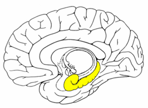
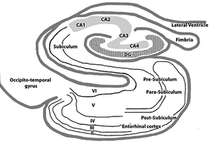
Hippocampus er beliggende mesialt i temporallappen og er funktionelt en del af det limbiske system (Fig. 1). Hippocampus udgør en skjult cortex, men en cortex med en simplere 1 cellelags opbydning (archicortex), sammenlignet med neocortex, hvor der typisk er 6 cellelag. Hippocampus spiller en afgørende rolle for indlæring og hukommelse, med særlig relation til sproglig hukommelse i dominant side (typisk venstre) og billedhukommelse i non-dominant side, ligesom Hippocampus spiller en rolle for evnen til spatiel orientering. Neurokirurgisk behandling i relation til Hippocampus er næsten udelukkende relateret til epilepsikirurgi og sjældnere gliomkirurgi. Hippocampus betyder søhest og har formodentligt fået dette navn pga. dens udseende i tværsnit. Man skal være opmærksom på om det blot er Hippocampus som anatomisk struktur der omtales, eller den Hippocampale formation som omfatter dele af Subiculum beliggende i overgangen mellem Hippocampus og gyrus parahippocampale, samt Entorhinale cortex beliggende i gyrus parahippocampale (Fig. 2).
Anatomi makroskopisk
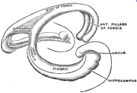
Hippocampus er beliggende mesialt i temporallappen og nås kirurgisk via temporalhornet af lateral ventriklen. Hippocampus danner mesialvæggen og gulvet af temporalhornet. Hippocampus opdeles i sin foreste del Pes Hippocampi, der lidt ligner en løvefod når den ses fra oven. Pes Hippocampus omtales også som Hippocampus hovedet, hvor demarkeringen mellem hovedet og kroppen af Hippocampus er Choroidal point. Choroidal point, er det sted hvor a. choroidea anterior træder ind i temporalhornet af lateral ventriklen gennem fissura choroidea. Den mesiale del af Hippocampus vender ind mod cisterna ambiens, hvor Hippocampus i anterior-posterior orientering buer sig rundt om lateralsiden af hjernestammen. Ved resektion af Hippocampus er arachnoidea ved cisterna ambiens den "sikre" mesiale grænse for resektionen, således at forstå at hvis denne arachnoidale "skillevæg" lades intakt, er essentielle strukturer som hjernestammen, kranienerver, og arterierne i cisterna ambiens beskytte på den anden side af arachnoidea. Opad til, eller dorsalt, adskilles Hippocampus fra Insula & basalganglier af fissura choroidea. Fornix udgør den dorsomediale del af Hippocampus kaldet Fimbria (Fig.3)
Anatomi mikroskopisk
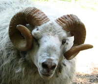
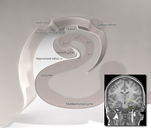
Funktionelt er Hippocampus tilsyneladende primært fungerende i kredsløb der er transverselt orienteret (Fig. 2). Hippocampus er som overfor omtalt sammenlignet med en søhest, men i tværsnittet er Hippocampus blev af den danske anatom Jacob Winsløw i 1732 sammenlignet med et bukkehorn (Ram's horn) på grund af den snoede form (Fig. 4). Senere blev strukturen af en fransk kirurg (de Garengeot) sammenlignet med den egyptiske gud Amuns horn (Cornu Ammonis), som har lagt navm til underindelingen af Hippocampus i CA1, CA2, CA3 & CA4. CA1-4 omtales ofte som den egentlige Hippocampus (Hippocampus proper), mens disse strukturer sammen med Gyrus dentatus, subiculum og entorhinale cortex omtales som den Hippocampale formation.
Det hippocampale kredsløb
Gyrus Dentatus består af tæt pakkede granulære celler som ligger henover CA4 - CA3 - CA2 - CA1. CA-delene af "hornet" består af tætpakkede pyramide celler svarende til dem man finder i neocortex. Efter CA1 kommer subiculum beliggende i den mesiale del af gyrus parahippocampale. Subiculum er bindeleddet til den Entorhinale cortex, som således har bragt os til den inferiore del af gyrus parahippocampale.
Det primære input til Hippocampus kommer fra den Entorhinale cortex, med signalprocessering frem mod nucleus dentatus. Output strukturen går fra Gyrus dentatus til subiculum. Dette er således grundstrukturen i det segmentale kredsløb i Hippocampus. Der er også kommunikation på langs af Hippocampus, men betydningen af disse baner er ikke afklaret. I enkelte mindre studier har det været foresået at kirurgisk opdeling af Hippocampus med tværgående snit som respektere det segmentale kredsløb, men med deling af de længdeforløbende fibre, kan være lige så effektivt som hippocampectomi ved epilepsikirurgi men med reduceret risiko for hukommelsespåvirkning. Der er også mange andre input til Hippocampus fra blandt andet: Amygdala, Claustrum, Substantia Innomata, Meynerts kerne, Thalamus, hypothalamus, retikulære formation i tegmentum, m.fl.
